The problem
Cancer is a leading cause of death worldwide. In particular, colorectal cancer is concerned with the large intestine (colon), the lower part of the digestive system, and the rectum (the last inches of the colon), and it is one of the most commonly diagnosed malignant neoplasms across genders. Even if colonoscopy is widely used for colorectal cancer detection and prevention, and it is considered nowadays as the gold standard for colon screening, there are still open challenges to overcome, such as the reduction of the polyps missing rate. A polyp is an abnormal growth of tissue projecting from a mucous membrane, and some types of polyps (e.g. adenoma) are premalignant conditions for colorectal cancer. Endoscopic resection during colonoscopy can prevent colorectal cancer development.
The main motivation for designing a computer-aided decision support system in colonoscopy is to assist a medical expert in improving the accuracy and promptness of medical diagnosis. It may also be used as an educational resource, speeding up the learning curve of medical students. One of the aims of developing a (semi)automatic classification system is to distinguish lesions which should be biopsied from those which should be directly removed during colonoscopy. A tool to perform virtual biopsy could be of great help to select lesions that must be directly resected (instead of performing biopsy), and thus avoid waiting for the histopathology to decide the therapeutic strategy. Furthermore, in a real clinical environment, virtual biopsy could help one to discriminate the severity of individual lesions in patients with many polyps, and the gastroenterologist could then focus on those requiring polypectomy.
Methods employed
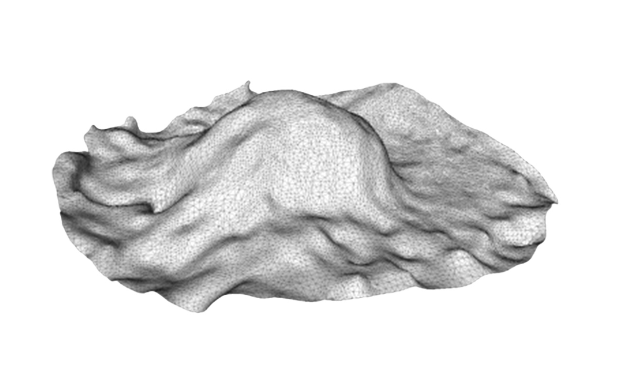
We used machine learning techniques (like support vector machines and random forest) to learn from data to discriminate the different types of lesions; and we employed computer vision techniques to both reconstruct the 3D shape of the lesions from the colonoscopy videos, and characterize each lesion at the level of color, texture and 3D shape.

Results
The work carried out by Dr. Pablo Mesejo, in collaboration with Dr. Daniel Pizarro and Dr. Adrien Bartoli, as well as with medical doctors Sylvain Beorchia, Laurent Poincloux, Olivier Rouquette, and Armand Abergel, resulted in a tool capable of improving the predictive accuracy of an expert gastroenterologist by almost 10%. This tool also saves clinician’s time by avoiding chromoendoscopy, a costly process (in temporal and economic terms) of colon staining with indigo carmine. The results of this research were published in one of the best journals in medical image analysis: Mesejo, P., Pizarro, D., Abergel, A., Rouquette, O., Beorchia, S., Poincloux, L., & Bartoli, A. (2016). Computer-aided classification of gastrointestinal lesions in regular colonoscopy. IEEE transactions on medical imaging, 35(9), 2051-2063. The software developed was registered on the pertinent French authority (IDDN.FR.001.350029.000.S.P.2015.000.31230. CAPRE (Computer-Aided Polyp Recognition), registered on 25/08/2015). The database compiled by Dr. Pablo Mesejo and Dr. Daniel Pizarro is available for public consultation at http://www.depeca.uah.es/colonoscopy_dataset/ and https://archive.ics.uci.edu/ml/datasets/Gastrointestinal+Lesions+in+Regular+Colonoscopy
Participants
Institutions
This project belonged to a larger one involving 5 French institutions through an ANR (French National Agency for Research) funded with 797.568€: SYSEO project (“Multimodal and Multimedia Image Analysis and Collaborative Networking for Digestive Endoscopy”).
Researchers
Dr. Pablo Mesejo as postdoctoral researcher in the ALCoV (Advanced Laparoscopy and Computer Vision) team, led by Dr. Adrien Bartoli, belonging to the ISIT laboratory (Image Sciences for Interventional Techniques, UMR 6284 – CNRS) at the Faculty of Medicine of the University of Auvergne Clermont-Ferrand I (France).